Neuromonitoring Carotid Endarterectomy

In the last post, I talked about an email asking for information about neuromonitoring for carotid endarterectomy surgeries. I told the emailer… “No, problem. Maybe I’ll even put a presentation together that will help you and other people reading this blog.”

Instead of posting the info like I was asked, I went on a rant about using motor evoked potentials for CEA cases.

neuromonitoring carotid endarterectomy rant

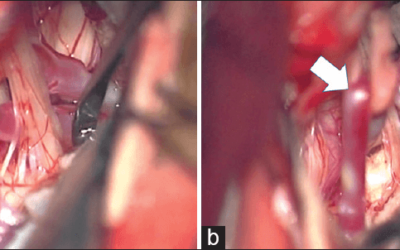
Image from http://goo.gl/q6ruJ

There was even some further discussion that went on about it on my linkedin account (just scroll down and look for my post about it).

Now, about 3-4 weeks later, I finally put together some presentation material that should help out, or at least give you a head start. It took way longer than I had planned, so if you find this useful, please let me know. Either leave a comment, scroll down and rate it 10 stars, post it somewhere like facebook or linkedin, etc. If enough people find it useful, then I might do some more in the future. If not, then there is no reason for me to put in the time that I did.

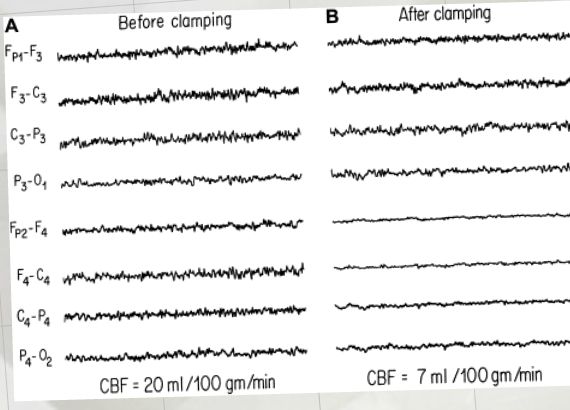
Anyways, the request was to help them make a presentation for vascular surgeon to try to drum up some business. They wanted to let them know the benefits of neuromonitoring carotid endarterectomy procedures. While other modalities like transcranial doppler, cerebral oximetry and stump pressure are commonly used, the request was for EEG and SSEP.

So I broke it down into three sections.

  1. What is the basis of neuromonitoring  for carotid endarterectomy procedures (using EEG and SSEP), and how does it work
  2. Is it of benefit
  3. Examples of how using neuromonitoring during CEA can assist the surgeon that is impossible without it

There’s plenty of other ways to present this, so here’s a couple templates for you to play with.

Here’s a scribd presentation…

Neuromonitoring Carotid Endarterectomy


Here’s a video for monitoring carotid endarterectomy surgeries…


Here’s my prezi for IONM CEA cases…


And here’s the transcripts to the prezi above…

A Closer Look Intraoperative Monitoring For Carotid Endarterectomy Start Here! Why IONM Is Effective 3 Points To Discuss Neuromonitoring during CEA is the more commonly used than any other cerebrovascular procedure. A survey performed by Chung et al. 1997 showed that almost 90% of CEA cases had some form of intraoperative neuromonitoring. Electroencephalogram (EEG) is considered the Gold Standard for monitoring CEA. Somatosensory Evoked Potentials (SSEP) are another commonly used modality to monitor electrical activity of the nervous system Both EEG and SSEP are used to monitor the electrical activity of the brain before, during and after clamping of the carotid artery. In order for EEG and SSEP to be successful at preventing stroke, there must be some relationship between the brain’s electrical activity and damage to brain matter. Identifying IONM Changes Does IONM Reduces Stroke Rates In CEA? – Risk of iatrogenic problems associated with shunting range from 0.5% to 3% (Sundt et al. 1986, Ahn and Concepcion 1995) The solution is selective shunting by the use of neuromonitoring. Why IONM Is Effective Does IONM Reduces Stroke Rates In CEA? Examples How You Can Use IONM To Reduce Patient Risk 1) Technical problems that limit the surgeon’s ability to expose and dissect the arothema, espcially the distal segment 2) Shunt kinking or occlusion due to improper placement 3) Intraoperative thrombosis 4) Increased risk of cerebral embolization of atherosclerotic debris and air into the distal circulation 5) Potential intimal damage resulting in postoperative thrombosis at the operative site. What does the EEG tell us: – The EEG reflects metabolic activity of the brain, which requires energy. Problems or alterations with energy production (increased demand or reduced supply) by brain cells can profoundly affect EEG activity. – EEG is a window into the functional and structural integrity of the brain. Some studies have shown SSEP to have remarkably higher sensitivity and specificity compared to EEG (Lam et al 1991, Haupt and Horcsh 1992). This could be due to the fact that SSEP also monitor subcortical structures (like the M1 branch to the basal ganglia, as well as the M2 branch Parietal lobe). – Significant changes seen with neuromonitoring happens about every 1 out of 10 cases, eliminating the risk associated with shunting in about 90% of CEA cases. – Selective shunting based on neuromonitoring, particualy major EEG changes, may reduce the incidence of stroke 10-fold (Newer, 1993), – Studies have shown neuromonitoring of CEA to even come “close to an irreducible minimum” (1.1% and 0%, Cho et al. 1986 and Bloom et al. 1990) – Woodworth et al 2007 reported a multivariate outcome analysis of selective versus routine shunting during 1411 CEAs. – Two out of 194 patients (1%) in the selective shunting group (based on EEG/SSEP) had perioperative strokescompared to 47 out of 1217 (4%) in the routine shuntinggroup. – Patients who had EEG monitoring with selective shunting were more than SEVEN X’s less likely to experience a perioperative stroke. (Salvian et al. 1997) compared the results of routinely shunted patients in 92 CEAs with selectively shunted patients in 213 CEAs (based on EEG monitoring). In the selectively shunted group, 16% subsequently required shunting. The major stroke rate in the routinely shunted group was 4.4%, and 0.5% in the selectively shunted group Using the bipolar montage and alarm criteria as outlined by Craft 1994, changes in EEG yielded a 100% sensitivity and 100% specificity for detection of an ischemic episode. Changes in EEG patterns have been shown to demonstrate ischemia to different brain tissue type, with a loss of fast rhythms associated with gray matter and an increase in slow rhythms reflects the deafferentation of the cortex caused by white matter ischemia (Faught, 1993) – General ischemia is seen at a cerebral blood flow of 10 ml/100g/min. – Tissue receiving flow between 18 and 23 ml/100g/min is functionally inactive, but function can be restored at any time with the reinstituion of increased perfusion (penlucida). – At lower levels, infarction is a function of time. If it recovers in time, no tissue damage happen (penumbra). (Young and Cole, 1993) Testing With Regional Cerebral Blood Flow (rCBF) Penlucida Changes in IONM v.s. Changes in Cerebral Blood Flow Brain Ischemia SSEP EEG – Whereas neuronal function is impaired immediately when blood flow drops below the threshold, the development of irreversible morphological damage is time dependent. Of course, once morphological damage becomes apparent, the initially reversible functional deficit turns into a persistent defect. – In order to PREVENT persistent defects, neuromonitoring must be sensitive enough to identify these problems early. Along the lines of nonfunctioning, noninfarcted, physically intact neurons, these are at a level of blood flow that will allow them to recover even if the intervention fails. The penumbra area is time dependant, while the penlucida is not. The rCBF levels are 18-23 ml/100g/min. – rCBF starts to show cortical decline at about 18-20 ml/100g/min and there will be a decrease in SSEP amplitude. – At 18 ml/100g/min, fast frequencies activity will be lost in EEGs. – At 15 ml/100g/min, there will be an increase in slow frequencies activity and loss of postsynaptic evoked potentials. At this level, synaptic activity decreases and you will see loss of brain electrical activity. – At 12 ml/100g/min you will see isoelectric EEG and loss of postsynaptic evoked potentials. At these levels, neuronal damage will occur if not changed.


Keep Learning

Here are some related guides and posts that you might enjoy next.

Intraoperative EMG: Referential or Bipolar?

Intraoperative EMG: Referential or Bipolar?

Recording Electrodes For EMG in the Operating Room: Referential or Bipolar? If your IONM manager walked into the OR in the middle of your case, took a look at your intraoperative EMG traces and started questioning your setup, could you defend yourself? I try to do...

read more
BAER During MVD Surgery: A New Protocol?

BAER During MVD Surgery: A New Protocol?

BAER (Brainstem Auditory Evoked Potentials) During Microvascular Decompression Surgery You might remember when I was complaining about using ABR in the operating room and how to adjust the click polarity to help obtain a more reliable BAER. But my first gripe, having...

read more

Bye-Bye Neuromonitoring Forum

Goodbye To The Neuromonitoring Forum One area of the website that I thought had the most potential to be an asset for the IONM community was the neuromonitoring forum. But it has been several months now and it is still a complete ghost town. I'm honestly not too...

read more

Pin It on Pinterest

Share This