Cranial Nerve Monitoring… Continued
In the last post, Cranial and Peripheral Nerve Monitoring – Clinical Uses Part 1, I went over a scenario where an unanticipated postoperative problem occurred due to poor neuromonitoring. The SNP’s feedback led the surgeon to believe the opposite of what was true. It doesn’t matter if you’re doing peripheral cranial nerve monitoring or central cord monitoring, poor neuromonitoring is probably worse than no monitoring at all.
Now back to our story…
Scratching your head, you figure out that today wasn’t the shining moment in your IOM career. Here’s what you had to offer:
- False negative EMG on the first side, since the nerve was cut and you didn’t get any activity.
- False positive tEMG on the first and second sides (ok, debate if you want to label this as a real false positive or not since he didn’t stimulate the whole nerve), since the surgeon only stimulated distal to the lesion to get a response.
- True positive EMG on the second side, since the patient woke up with an injury on that side.
Even though your EMG was correct on the second side, it missed the severing of the nerve on the first side. And in this case, we aren’t sure as to what extent it helped in preventing injury. For the second side, we’ll never know if it was the initial insult, the second, or a compounding effect. EMG does a poor job at qualifying the extent of the injury.
You also realize that the disappearance of train EMG and stimulating the portion of the nerve distal to the operative site offers poor clinical data.
What’s The Peripheral and Cranial Nerve Monitoring Lesson Learned?
It’s very likely that the surgeon severed the nerve on the first side, and there was not enough activity for the EMG to pick up a response (reports of this are seen in the literature as well). So if the surgeon stimulated the proximal and distal nerve on the first side, he would have realized that the nerve was severed completely.
The surgeon might not have risked injury to the other nerve since vocal paresis is not as detrimental as vocal paralysis. He might have second-guessed his opinion to aggressively remove the tumor on the second side with a severed nerve on the other and train EMG on the second side. Most, in my experience, will stage the procedure at this point. That means figuring out what else to do, if anything, on the first side, and then close up. Give the patient some time for assessment and resechedule the second side at a later date.
A second thought is to be more active with the probe during exposure and prior to and during tumor debulking. Mapping out the nerve might have led to a better outcome, especially if the tumor made visualizing the nerve difficult.
While I made this whole scenario up, it’s not far-fetched to find yourself in this very predicament. The best way to handle something like this is to prevent it before it happens in the first place.
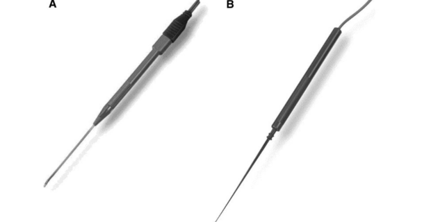
- Let the surgeon know what the best stimulating techniques are for identifying a cranial nerve.
- Talk during the surgery so you can better interpret your free-running EMG by knowing what the surgeon is doing.
- Inform the surgeon of the strengths and weaknesses of your modalities.
- Be better than the automated system.
Anyone else ever has a case similar while performing cranial nerve monitoring? What was done differently, or the same?
Keep Learning
Here are some related guides and posts that you might enjoy next.
How To Have Deep Dive Neuromonitoring Conversations That Pays Off…
How To Have A Neuromonitoring Discussion One of the reasons for starting this website was to make sure I was part of the neuromonitoring conversation. It was a decision I made early in my career... and I'm glad I did. Hearing the different perspectives and experiences...
Intraoperative EMG: Referential or Bipolar?
Recording Electrodes For EMG in the Operating Room: Referential or Bipolar? If your IONM manager walked into the OR in the middle of your case, took a look at your intraoperative EMG traces and started questioning your setup, could you defend yourself? I try to do...
BAER During MVD Surgery: A New Protocol?
BAER (Brainstem Auditory Evoked Potentials) During Microvascular Decompression Surgery You might remember when I was complaining about using ABR in the operating room and how to adjust the click polarity to help obtain a more reliable BAER. But my first gripe, having...
Bye-Bye Neuromonitoring Forum
Goodbye To The Neuromonitoring Forum One area of the website that I thought had the most potential to be an asset for the IONM community was the neuromonitoring forum. But it has been several months now and it is still a complete ghost town. I'm honestly not too...
EMG Nerve Monitoring During Minimally Invasive Fusion of the Sacroiliac Joint
Minimally Invasive Fusion of the Sacroiliac Joint Using EMG Nerve Monitoring EMG nerve monitoring in lumbar surgery makes up a large percentage of cases monitored every year. Using EMG nerve monitoring during SI joint fusions seems to be less utilized, even though the...
Physical Exam Scope Of Practice For The Surgical Neurophysiologist
SNP's Performing A Physical Exam: Who Should Do It And Who Shouldn't... Before any case is monitored, all pertinent patient history, signs, symptoms, physical exam findings and diagnostics should be gathered, documented and relayed to any oversight physician that may...

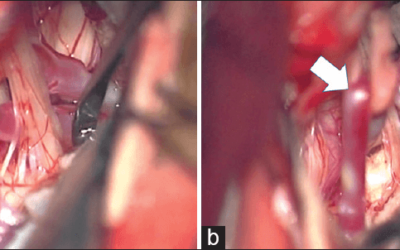


Had a case where the surgeon severed the nerve. He knew it, and stimulation proved it. He still went ahead and did the other side.