Glioma Surgery… Is Intraoperative Monitoring Wort It?
Glioma tumors in the brain (which is a common malignant brain tumor) provides a intraoperative monitoring conundrum for the surgeon.
Typically, can we offer the surgeon the ability to better localize the central sulcus (through phase reversal), monitor the integrity of the sensory system (through somatosensory evoked potentials) and monitor the integrity of the motor system (through direct cortical motor evoked potentials).
This should help reduce morbidity rates by preventing the neurological system from injury. However, the mortality rates of patient’s with a glioma tumor in the brain are directly related to the amount of tumor removed. The less tumor they take out, the less lifespan they have.
If the surgeon decides to stop resecting the tumor because the neuromonitoring team gave a warning of injury (doesn’t matter if done correctly or not), the patient may not have as long to live. If the surgeon forgoes intraoperative monitoring, the patient might be at higher risk of neural compromise along with that extended life.
Intraoperative monitoring came on the scene out of necessity. Preoperative imaging as a predictor of function through classic anatomical criteria was found to be insufficient. This was due to variability of cortical organization, distortion of the cerebral topography as a result of mass effect of the tumor, the functional reorganization because of plasticity and shift of cortical and subcortical fibers throughout the procedure that makes preoperative tractography inaccurate. Intraoperative monitoring was brought on to help with intraoperative localization and monitor functional integrity.
Phase Reversal
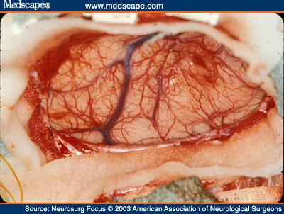
Phase reversal was first described by Goldring (1978), and then later used for tumor surgery. This modality has worked well for localization, better serving the surgeon to identify important cortical structures and orient him/herself.

Image from http://bit.ly/190dsY6
Direct Motor Evoked Potentials
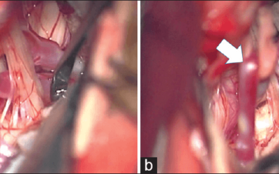
Taniguchi (1993) first described a modification of monopolar stimulation, which would later be adapted to a bipolar stimulation that would demonstrate a correlation between intraoperative CMAP changes and postoperative clinical outcome. Although there seems to be clinical relevance for glioma surgeries, there has been criticism that IOM leads to less tumor resection. Others (Kombos, 2009) have reported that there is no negative influence of IOM with amount of tumor resection. They found survival rates to be the same in both intraoperative monitored groups and those without, while improving the safety of the procedure.

Image from http://bit.ly/1648V5X
Intraoperative Tractography
In a study performed by Maesawa (2010), they found that intraoperative tractography demonstrated the location of the corticospinal tract more accurately than preoperative tractography. This enabled them to safely approach the corticospinal tract, while removing more tumor.
Real time intraoperative tractography
Of interest to us, they used the intraoperative tractography along with subcortical MEP monitoring in their identification of the corticospinal tracts (along with phase reversal and cortical motor evoked potentials on the primary motor cortex).
The subcortical MEP intensity was set to that of 50% of a reliable direct cortical MEP. This is due to the fact that the subcortical MEP is directly stimulating the white tracts of the corticospinal tract. They would go up to 200% of the direct cortical MEP stimulation level, and never over 30mA.
Combined, these 2 modalities found that shift of these tracts was up to 23mm inward and 12 mm outward, with an absolute value of the shift at 6.65 mm. They used the MEP to help identify the distance by comparing the intensity to threshold to the intraoperative corticospinal tract tractography. There was a direct relationship. This was not the same when using MEP and pre-operative tractography, which had no significant correlation at all.
They were able to identify that if subcortical stimulation was 100% of direct cortical stimulation of MEP (or exactly the same level), then the distance was about 7 mm to the tract. This distance (as correlated by intensity level) allowed them to approach the corticospinal tracts with an estimate as to how much room they have to work with. This allowed for safer tumor removal.
What, You Don’t Have All The Toys?
Me either.
But that’s OK. Though a bit controversial, there are surgeons that do value intraoperative monitoring of glioma tumors as it’s typically performed.
Being able to better localize the central sulcus, and have an idea as to how the primary motor strip is functioning, can be useful information. If the surgeon is aware of the strengths and weakness of the monitoring, s/he should be able to make informed decisions and provide the best care possible for that patient.
Keep Learning
Here are some related guides and posts that you might enjoy next.
How To Have Deep Dive Neuromonitoring Conversations That Pays Off…
How To Have A Neuromonitoring Discussion One of the reasons for starting this website was to make sure I was part of the neuromonitoring conversation. It was a decision I made early in my career... and I'm glad I did. Hearing the different perspectives and experiences...
Intraoperative EMG: Referential or Bipolar?
Recording Electrodes For EMG in the Operating Room: Referential or Bipolar? If your IONM manager walked into the OR in the middle of your case, took a look at your intraoperative EMG traces and started questioning your setup, could you defend yourself? I try to do...
BAER During MVD Surgery: A New Protocol?
BAER (Brainstem Auditory Evoked Potentials) During Microvascular Decompression Surgery You might remember when I was complaining about using ABR in the operating room and how to adjust the click polarity to help obtain a more reliable BAER. But my first gripe, having...
Bye-Bye Neuromonitoring Forum
Goodbye To The Neuromonitoring Forum One area of the website that I thought had the most potential to be an asset for the IONM community was the neuromonitoring forum. But it has been several months now and it is still a complete ghost town. I'm honestly not too...
EMG Nerve Monitoring During Minimally Invasive Fusion of the Sacroiliac Joint
Minimally Invasive Fusion of the Sacroiliac Joint Using EMG Nerve Monitoring EMG nerve monitoring in lumbar surgery makes up a large percentage of cases monitored every year. Using EMG nerve monitoring during SI joint fusions seems to be less utilized, even though the...
Physical Exam Scope Of Practice For The Surgical Neurophysiologist
SNP's Performing A Physical Exam: Who Should Do It And Who Shouldn't... Before any case is monitored, all pertinent patient history, signs, symptoms, physical exam findings and diagnostics should be gathered, documented and relayed to any oversight physician that may...


