I came across some papers on motor evoked potentials (MEPs) that got me thinking a little bit.
It got me thinking about small influences in the operating room that may, or may not, affect our tracings during our cases. Specifically, MEPs.
Here’re some snippets from the 2 articles:
Kamibayashi K (2009)
Specifically, to investigate the effect of load-related afferent inputs on the corticospinal excitability during passive stepping, motor evoked potentials (MEPs) in response to the stimulation were compared between two passive stepping conditions: 40% body weight unloading on a treadmill (ground stepping) and 100% body weight unloading in the air (air stepping). In the rectus femoris, biceps femoris and tibialis anterior (TA) muscles, electromyographic activity was not observed throughout the step cycle in either stepping condition. However, the TMS-evoked MEPs of the TA muscle at the early- and late-swing phases as well as at the early-stance phase during ground stepping were significantly larger than those observed during air stepping.
and Kitamura T (2012)
During passive stepping, the MEP amplitudes in FCR muscle were significantly increased in six adjacent stimulus sites of the hot spot
For chiropractors, PT’s, OT’s, etc that do physical rehab, these findings should come at no shock. Afferent bombardments are used all the time to improve neurological function. We utilize the pain gate theory by using TENS units to stimulate below pain thresholds (larger diameter afferent fibers) to inhibit the slower pain fibers (smaller diameter afferents) at the spinal and cortical levels. Chiropractors will adjust the spine to change the gain of muscle spindle fibers and restore segmental motion, thereby creating a heightened level of afferent feedback.
This kind of stuff is really old hat.
MEPs in the OR
But it got me thinking about those very fickle motor evoked potentials that we perform on an anesthetized patient in the operating room. What kinds of things would influence MEPs in that environment? Well here’s some of the much-studied causes:
In addition, here’s a short (incomplete) list of commonly accepted factors:
- Anesthesia
- Blood pressure
- Recording and stimulating electrode placement
- Case duration/Fade
- Blood loss
- Patient temperature
But according to Kamibayashi K (2009) and Kitamura T (2012), there can be an increase in the amplitude of muscles with afferent stimulation. And this can happen to the muscle being loaded, like the tibialis anterior in the weighted portion of normal gait, as well as a distant muscle, like the arm flexors during weighted gait.
And I’m aware of studies like Journee (2007) Léonard G (2012), and Lapole T (2012) where they’ve experimented with front loading MEPs with afferent stimulation and applying a double stimulation to utilize temporal facilitation.
But what I’m wondering about is the intermittent afferent stimuli affecting MEPs that is out of our control…
- Do MEPs improve when the blood pressure cuff turns on? Is it significant?
- What about the lower extremities with cases with intermittent sequential pneumatic compression? Do those make a difference?
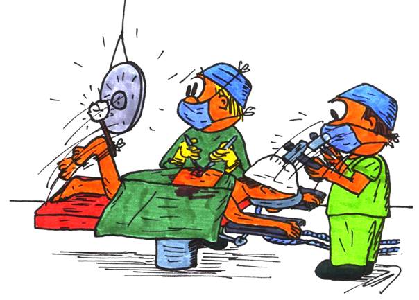
- Is there any ramped up feedback when the patient is forced into dorsiflexion at the ankle on certain tables? Does it last consistently?
- How about if the patient is getting low levels of anesthesia and begins reacting to pain, as seen on EMGs. I wouldn’t want to shock them then, but I wonder what their MEPs look like then?
Like I said, this was just something I was wondering about. I’m not sure if my thought process was of any benefit, but maybe those were some papers you were unaware of.
What about you, ever wonder about MEPs?
Keep Learning
Here are some related guides and posts that you might enjoy next.
How To Have Deep Dive Neuromonitoring Conversations That Pays Off…
How To Have A Neuromonitoring Discussion One of the reasons for starting this website was to make sure I was part of the neuromonitoring conversation. It was a decision I made early in my career... and I'm glad I did. Hearing the different perspectives and experiences...
Intraoperative EMG: Referential or Bipolar?
Recording Electrodes For EMG in the Operating Room: Referential or Bipolar? If your IONM manager walked into the OR in the middle of your case, took a look at your intraoperative EMG traces and started questioning your setup, could you defend yourself? I try to do...
BAER During MVD Surgery: A New Protocol?
BAER (Brainstem Auditory Evoked Potentials) During Microvascular Decompression Surgery You might remember when I was complaining about using ABR in the operating room and how to adjust the click polarity to help obtain a more reliable BAER. But my first gripe, having...
Bye-Bye Neuromonitoring Forum
Goodbye To The Neuromonitoring Forum One area of the website that I thought had the most potential to be an asset for the IONM community was the neuromonitoring forum. But it has been several months now and it is still a complete ghost town. I'm honestly not too...
EMG Nerve Monitoring During Minimally Invasive Fusion of the Sacroiliac Joint
Minimally Invasive Fusion of the Sacroiliac Joint Using EMG Nerve Monitoring EMG nerve monitoring in lumbar surgery makes up a large percentage of cases monitored every year. Using EMG nerve monitoring during SI joint fusions seems to be less utilized, even though the...
Physical Exam Scope Of Practice For The Surgical Neurophysiologist
SNP's Performing A Physical Exam: Who Should Do It And Who Shouldn't... Before any case is monitored, all pertinent patient history, signs, symptoms, physical exam findings and diagnostics should be gathered, documented and relayed to any oversight physician that may...


